Many of the test results (as described in the Diagnosis section) are used to determine the extent or stage of the cancer. The stage of cancer describes how much cancer is in the body. It helps your doctor understand the seriousness of the cancer, if and where it has spread, and how best to treat it. From this information, the oncologist can also assess the likelihood of survival.
For cancer of the larynx, doctors mostly depend on the TNM system created by the American Joint Committee on Cancer (AJCC). The TNM system is based on 3 key pieces of information:
- How big the main tumor (T) is
- If the cancer has spread to nearby lymph nodes (N)
- The spread (metastasis) to distant parts of the body (M)
Numbers or letters after T, N, and M provide more details about each of these factors. Lower numbers mean that the cancer is in an early stage. Higher numbers mean the cancer is more advanced.
Tumor (T)
- TX: The primary tumor cannot be evaluated.
- Tis: This is a stage called carcinoma (cancer) in situ. It is a very early cancer where cancer cells are found in only 1 layer of tissue.
When describing T1 to T4 tumors, doctors divide the larynx into 3 regions: the supraglottis, glottis, and subglottis.
Knowing the three main parts of the larynx will help you understand the laryngeal cancer stages and grades.
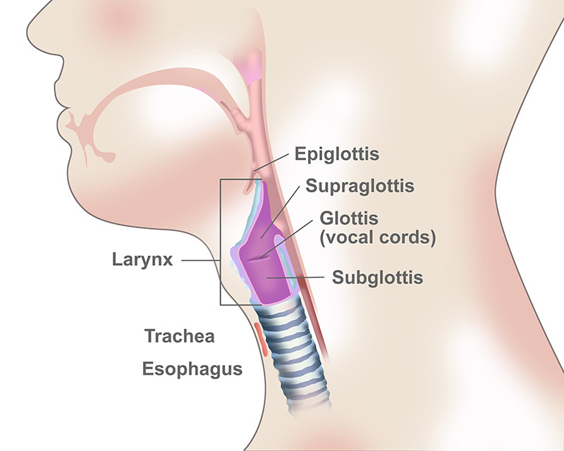
Supraglottis tumor of the larynx
- T1: The tumor is located in a single area above the vocal cords that does not affect the movement of the vocal cords.
- T2: The tumor started in the supraglottis, but it has spread to the mucous membranes that line other nearby areas, such as the base of the tongue. The vocal cords are not affected.
- T3: The tumor is limited to the larynx and affects the vocal cords. The tumor may have spread to surrounding tissue.
- T4: The tumor has spread beyond the larynx.
- T4a: The tumor has spread through the thyroid cartilage and/or the tissue beyond the larynx.
- T4b: The tumor has spread to the area in front of the spine (prevertebral space) or the chest area, or it encases the arteries.
Glottis tumor of the larynx
- T1: The tumor is limited to the vocal cords, but it does not affect the movement of the cords.
- T1a: The tumor is only in the right or left vocal cord.
- T1b: The tumor is in both vocal folds.
- T2: The tumor has spread to the supraglottis and/or the subglottis. The tumor may also affect the movement of the vocal cord.
- T3: The tumor is limited to the larynx and paralyzes at least 1 of the vocal cords. The tumor may also invade the space inside the larynx and/or the cartilage around the thyroid gland.
- T4: The tumor has spread beyond the larynx.
- T4a: The tumor has spread to the thyroid cartilage and/or the tissue beyond the larynx.
- T4b: The tumor has spread to the area in front of the spine (prevertebral space) or the chest area, or it encases the arteries.
Subglottis tumor of the larynx
- T1: The tumor is only in the subglottis.
- T2: The tumor has spread to the vocal cords. Movement of the vocal cords may be affected.
- T3: The tumor is limited to the larynx and affects the vocal folds. It may also invade the space inside the larynx and/or the cartilage of the thyroid.
- T4: The tumor has spread beyond the larynx.
- T4a: The tumor has spread to the cricoids, the ring-shaped cartilage near the bottom of the larynx, or thyroid cartilage and/or the tissue beyond the larynx.
- T4b: The tumor has spread to the area in front of the spine or the chest area, or it encases the arteries.
Node (N)
Evaluation of nodes can be clinical or pathological.
Clinical N
- NX: The regional lymph nodes cannot be evaluated.
- N0 (N plus zero): There is no evidence of cancer in the regional lymph nodes.
- N1: The cancer has spread to a single lymph node on the same side as the primary tumor, and the cancer found in the node is 3 cm or smaller. There is no ENE.
- N2a: Cancer has spread to a single lymph node on the same side as the primary tumor and is larger than 3 cm but not larger than 6 cm. There is no ENE.
- N2b: Cancer has spread to more than 1 lymph node on the same side as the primary tumor, and none measures larger than 6 cm. There is no ENE.
- N2c: Cancer has spread to more than 1 lymph node on either side of the body, and none measures larger than 6 cm. There is no ENE.
- N3a: The cancer is found in a lymph node and is larger than 6 cm. There is no ENE.
- N3b: There is ENE in any lymph node.
Pathological N
- NX: The regional lymph nodes cannot be evaluated.
- N0 (N plus zero): There is no evidence of cancer in the regional lymph nodes.
- N1: The cancer has spread to a single lymph node on the same side as the primary tumor, and the cancer found in the node is 3 cm or smaller. There is no ENE.
- N2a: Cancer has spread to 1 lymph node and is 3 cm or smaller, but there is ENE. Or, cancer has spread to a single lymph node on the same side as the primary tumor and is larger than 3 cm but not larger than 6 cm, and there is no ENE.
- N2b: Cancer has spread to more than 1 lymph node on the same side as the primary tumor, and none measures larger than 6 cm. There is no ENE.
- N2c: Cancer has spread to more than 1 lymph node on either side of the body, and none measures larger than 6 cm. There is no ENE.
- N3a: The cancer is found in a lymph node and is larger than 6 cm. There is no ENE.
- N3b: There is ENE in a single lymph node on the same side as the primary tumor, and it is larger than 3 cm. Or, cancer has spread to many lymph nodes, and at least 1 has ENE. Or, there is ENE in a single lymph node on the opposite side of the primary tumor that is 3 cm or smaller.
Metastasis (M)
The "M" in the TNM system describes whether the cancer has spread to other parts of the body, called distant metastasis.
- M0: The cancer has not spread to other parts of the body.
- M1: The cancer has spread to other parts of the body.
Laryngeal Cancer Stage Grouping
Once the T, N, and M categories of the cancer have been determined, the information is combined in a process called stage grouping, which helps the doctor to assign an overall stage. The standardized stages of laryngeal cancer are:
- Stage 0 Laryngeal Cancer: abnormal cells in the top layer of cells lining of the larynx that may become cancer
- Stage I Laryngeal Cancer: cancer has grown deeper, but is only in one part of the supraglottis, and the vocal cords move normally
- Stage II Laryngeal Cancer: cancer has grown deeper and spread into more than one part of the supraglottis (or glottis), and the vocal cords move normally; it has not spread to nearby lymph nodes or to distant parts of the body
- Stage III Laryngeal Cancer: tumor is only in the larynx but has caused a vocal cord to stop moving, or the tumor is growing into nearby areas such as the postcricoid, paraglottic space, pre-epiglottic (in front of the epiglottis) tissues, or the inner part of the thyroid cartilage (firm tissue that separates the thyroid gland from the front of the larynx); the cancer has spread to a single lymph node on the same side of the neck and is no larger than 3 centimeters
- Stage IVA Laryngeal Cancer: cancer has spread to the cartilage around the thyroid or trachea, bone under the tongue, the thyroid, or nearby soft tissue (this is also known as moderately advanced local disease); it may or may not have spread to a single lymph node on the same side of the neck
- Stage IVB Laryngeal Cancer: cancer spread to the muscles in the upper spinal column, carotid artery, chest cavity lining, and/or lymph nodes (this is also known as very advanced local disease)
- Stage IVC Laryngeal Cancer: any size tumor has spread to other parts of the body
Grade (G)
Doctors also describe cancer of the larynx by its grade (G). The grade describes how much cancer cells look like healthy cells when viewed under a microscope.
Cancer that looks similar to healthy tissue and has different cell groupings is called a "differentiated" or "low-grade tumor." Cancer that looks very different from healthy tissue is called "poorly differentiated" or a "high-grade tumor." The cancer’s grade can help the doctor predict how quickly the cancer will spread. Generally, the lower the grade, the better the prognosis.
- GX: The grade cannot be evaluated
- G1: The cells look more like normal tissue (well differentiated)
- G2: The cells are moderately differentiated
- G3: The cells don’t resemble healthy tissue (poorly differentiated)
Head and Neck Cancer Care Available in South Chicago
If you have been diagnosed with cancer of the larynx or a different type of head and neck cancer, Affiliated Oncologists are ready to help. The comprehensive approach offered by our cancer team combines the latest laryngeal cancer treatments with education and support services. Our oncologists specialize in cancer care and are ready to talk to you about your diagnosis and personalized treatment options. Our cancer centers are located in Chicago Ridge, Mokena, Hazel Crest, Palos Heights, and Oak Lawn.