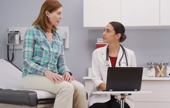
Doctors use many tests to examine the throat and neck in order to detect and diagnose laryngeal cancer. In many cases, you will start with an Ear, Nose, Throat specialist (ENT) who will perform the exams and request further testing if there are signs indicating that a tumor is present. Some of the following tests or procedures may be run to make a determination.
- Physical exam of the throat: An exam in which the doctor feels for swollen lymph nodes in the neck and looks down the throat with a small, long-handled mirror to check for abnormal areas. The doctor will also check the insides of the cheeks and lips; the gums; the back, roof, and floor of the mouth, and the top, bottom, and sides of the tongue.
If the doctor suspects there could be cancer, a biopsy may be performed as it is the only sure way for the doctor to know if an area of the body has cancer. A biopsy requires a tissue sample be collected from the area of the body where cancer is suspected so that the cells can be tested to see if cancer is present. There are a few ways that the larynx tissue may be collected:
- Laryngoscopy: A procedure in which the doctor checks the larynx (voice box) with a mirror or a laryngoscope to check for abnormal areas. A laryngoscope is a thin, tube-like instrument with a light and a lens for viewing the inside of the throat and voice box. It may also have a tool to remove tissue samples, which are checked under a microscope for signs of cancer.
- Endoscopy: A procedure to look at organs and tissues inside the body, such as the throat, esophagus, and trachea to check for abnormal areas. An endoscope (a thin, lighted tube with a light and a lens for viewing) is inserted through an opening in the body, such as the mouth. A special tool on the endoscope may be used to remove samples of tissue.
- Panendoscopy: A procedure that combines laryngoscopy, esophagoscopy, and (at times) bronchoscopy. This lets the doctor thoroughly examine the entire area around the larynx and hypopharynx, including the esophagus (swallowing tube) and trachea (windpipe). While the patient is under general anesthesia, the doctor will thoroughly examine all of these areas to look for tumors and determine their size and if they’ve spread. A tissue sample can be collected for biopsy during this procedure as well.
Tests to Determine if the Cancer Has Spread Outside of the Larynx
If the biopsy showed cancer, it’s likely your doctor will run other tests, including imaging, to see if the cancer has spread and, if so, how far. Your doctor may request one or more of the following imaging scans:
- CT scan (CAT scan): A procedure that makes a series of detailed pictures of areas inside the body, taken from different angles. The pictures are made by a computer linked to an x-ray machine. A dye may be injected into a vein or swallowed to help the organs or tissues show up more clearly. This procedure is also called computed tomography, computerized tomography, or computerized axial tomography.
- MRI (magnetic resonance imaging): A procedure that uses a magnet, radio waves, and a computer to make a series of detailed pictures of areas inside the body. This procedure is also called nuclear magnetic resonance imaging (NMRI).
- PET scan (positron emission tomography scan): A procedure to find malignant tumor cells in the body. A small amount of radioactive glucose (sugar) is injected into a vein. The PET scanner rotates around the body and makes a picture of where glucose is being used in the body. Malignant tumor cells show up brighter in the picture because they are more active and take up more glucose than normal cells do. In some cases, the PET scan and CT scan may be used together. This is called a PET-CT.
- Bone scan: A procedure to check if there are rapidly dividing cells, such as cancer cells, in the bone. A very small amount of radioactive material is injected into a vein and travels through the bloodstream. The radioactive material collects in the bones with cancer and is detected by a scanner.
- Barium swallow: A series of x-rays of the esophagus and stomach. The patient drinks a liquid that contains barium (a silver-white metallic compound). The liquid coats the esophagus and stomach, and x-rays are taken. This procedure is also called an upper GI series.
Learn more about how oncologists determine the stage of laryngeal cancer.
Laryngeal Cancer Specialists in the South Chicago Suburbs
If you or a loved one have been diagnosed with cancer of the larynx or another type of head and neck cancer, the cancer specialists at Affiliated Oncologists are ready to help. We have cancer centers offering the latest treatments available throughout South Chicago, including Chicago Ridge, Mokena, Hazel Crest, Palos Heights, and Oak Lawn.